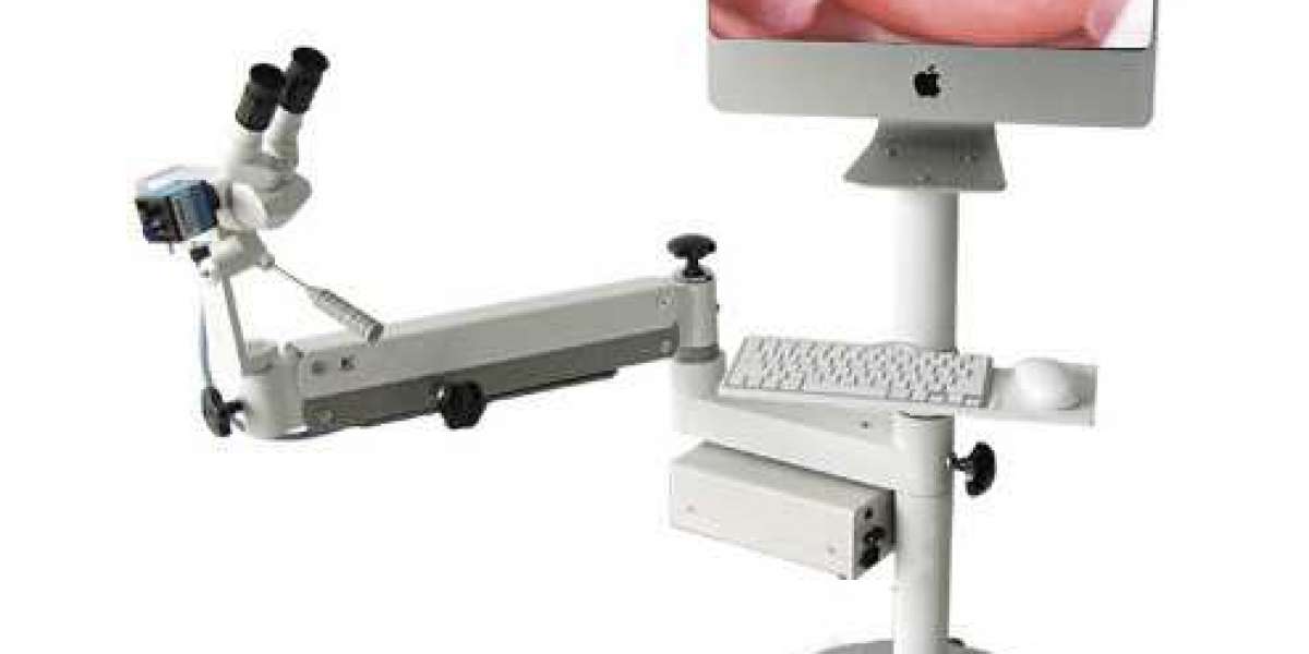
To demonstrate that colposcopy, which was first used in 1925, is still a valid technique that has not undergone significant modifications from the original approach described at the turn of the twentieth century, this review will focus on the following points:However, the Second World War, which occurred during the war, hampered the spread and development of colposcopy. Colposcopy has made significant advances in Spain, Italy, Brazil, France, and Switzerland, among other countries. When colposcopy was first introduced in the United States in the 1970s, it was primarily used by experts who had received extensive training in cytopathology, anatomic pathology, and colposcopy and who were able to diagnose and treat cervical abnormalities. Since then, it has become widely available.
Colposcopists were not trained as gynecologists in most European countries, in contrast to the situation in most other European countries, where colposcopists were trained as gynecologists and their histocytological expertise was slightly more broad than that of gynecologists. In countries with a strong Anglo-Saxon influence, colposcopy is only performed on a limited basis, whereas in countries with a strong German medical legacy, colposcopy is performed routinely during a standard general gynecological appointment. It should be noted that this distinction is not conclusive, and it does not imply that all European or Latin American gynecologists use colposcopy exclusively for training purposes, as has been suggested by some.
We developed dynamic colposcopy in 1977 to distinguish it from the descriptive immobility of Hinselmann's original classification (1954), which was largely unmodified by his immediate successors. This was done to distinguish it from the descriptive immobility of Hinselmann's original classification (1954). The goal of this project was to transform colposcopy gynecology into a diagnostic tool capable of identifying abnormal substrates in typical colposcopic images, which had previously been impossible. Using our findings, we were able to identify eight distinct markers that distinguish between ATZ areas that are secondary or non-biopsiable and those that are not.

As previously stated, the categorization method proposed in Rome (IFCPC, 1990) lends support to our initial hypothesis, as the ability to recognize large or minor alterations in the original photographs can aid in determining how severe damage caused by the explosion was. The specificity ranges from 48% to 10% of the population, while the sensitivity is 96%.
Clearly, for the method to be successful, a wide range of colposcopic specificity is required to be utilized. Should a biopsy that reveals only a low-grade lesion be regarded as a false positive colposcopic result, or should it be regarded as a benign lesion in the first place? However, even though histopathologic findings are considered the gold standard, it is widely acknowledged that there is some subjectivity in the assessment of these findings. When the same pathologist reviews the diagnosis after a period of time, there may be differences in the way the diagnosis is made between and within observers. A wide error range is believed to exist in microbiopsy performed under colposcopic control, which means that the results cannot be considered representative of the lesion under investigation. When untrained hands perform colposcopy-guided biopsy, or when biopsy is restricted to a small and insufficient sampling area, the likelihood of this occurring is increased.
When performing a complete visual assessment, it is common to overlook the squamocolumnar junction. Even if the severity of the endocervical lesions is in doubt, microbiopsies taken from the ectocervix cannot be used to assess their severity. Microbiopsies taken from the ectocervix cannot be used to assess the severity of any endocervical lesions. Endocervical involvement can be detected using a procedure known as microcolpohysteroscopy (MCH), which is performed using a small camera.
In light of the information presented here, colposcopy appears to be in good health, and its popularity in gynecology is likely to increase in the future if cytopathologists and gynecologists are not forced to act as makeshift specialists for their respective disciplines. When it comes to everyday practice, colposcopy should be considered standard, and the role of the gynecologist as the leading expert in integral women's health should be preserved.


